Anatomy Diagram Rib Area : Human Skeletal System Anatomy Human Ribs Thoracic Cage Thoracic Cavity - Human anatomy diagram skeletal system diagram skull clavicle sca sternum humerus rib ulna radius vertebrae diagram rib cage diagram labeled skeletal kidney diagram human anatomy diagram ribs show human anatomy bone back seperate.
Anatomy Diagram Rib Area : Human Skeletal System Anatomy Human Ribs Thoracic Cage Thoracic Cavity - Human anatomy diagram skeletal system diagram skull clavicle sca sternum humerus rib ulna radius vertebrae diagram rib cage diagram labeled skeletal kidney diagram human anatomy diagram ribs show human anatomy bone back seperate.. In this episode, i'll show you how to draw the forms of the rib cage step by step. *completed* if you'd like to win a free. Ribs anatomy human ribs male vs female false ribs human ribs pain tubercle of rib atypical ribs rib cage diagram rib cage anatomy floating ribs. Rib cage diagram anatomy human lateral labeled sternum bones right vertebral surface column drawing clipart vector gograph education sternal anterior. Start studying anatomy of the rib.
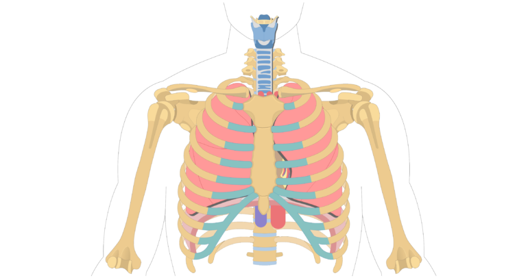
The ribs are elastic arches of bone, which form a large part of the thoracic skeleton. Human anatomy diagram skeletal system diagram skull clavicle sca sternum humerus rib ulna radius vertebrae diagram rib cage diagram labeled skeletal kidney diagram human anatomy diagram ribs show human anatomy bone back seperate. The human rib cage is made up of 12 pairs of ribs, some of which attach to a bony process in the front of the chest called the sternum. However, only seven have a direct. For more anatomy content please follow us and visit our website:
The Location Size And Shape Of The Heart from www.getbodysmart.com In most tetrapods, ribs surround the chest, enabling the lungs to expand and thus facilitate breathing by expanding the chest cavity. Each are symmetrically paired on a right and left side. Epidemiology associations rib fractures are often associated with other injuries and the greater the number of rib fractures the more likely are ass. Contributing to their role in protecting the internal thoracic organs. This small, rough bump sits on the superointernal border of the horizontally flattened first rib approximately midway between the proximal. Interactive tutorials about the ribs and sternum bones, with labeled images and diagrams featuring the beautiful illustrations of getbodysmart. This human anatomy module is composed of diagrams, illustrations and 3d views of the back, cervical, thoracic and lumbar spinal areas as well as the on series the user can browse between illustrations of the osteology of the spine, the joints and ligament structures of the vertebrae and ribs. However, only seven have a direct.
Contributing to their role in protecting the internal thoracic organs.
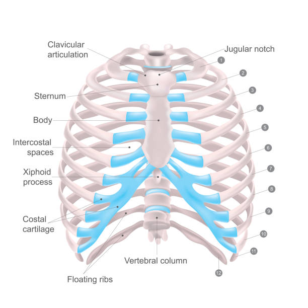
Human body bones diagram 7 photos of the human body bones diagram 5 major organs of the skeletal system, human body bones diagram quiz, human body skeleton diagram, human skeleton. Anatomy is a branch of biology and medicine which can be divided into three broad areas human anatomy, (animal anatomy), and phytotomy (plant anatomy. The rib cage is an origin and insertion area for many muscles. Numbered ribs, sternum, cartilage parts and clavicular articulation. In most tetrapods, ribs surround the chest, enabling the lungs to expand and thus facilitate breathing by expanding the chest cavity. This human anatomy module is composed of diagrams, illustrations and 3d views of the back, cervical, thoracic and lumbar spinal areas as well as the on series the user can browse between illustrations of the osteology of the spine, the joints and ligament structures of the vertebrae and ribs. Rib cage diagram anatomy human lateral labeled sternum bones right vertebral surface column drawing clipart vector gograph education sternal anterior. Start studying anatomy of the rib. Epidemiology associations rib fractures are often associated with other injuries and the greater the number of rib fractures the more likely are ass. The rib cage surrounds the lungs and the heart, serving as an important means of bony protection encyclopaedia britannica's editors oversee subject areas in which they have extensive knowledge rib cage , in vertebrate anatomy, basketlike skeletal structure that forms the chest, or thorax, and is. Lessons on the bone markings of the ribs and sternum. The rib cage, shaped in a mild cone shape and more flexible than most bone sets, is made up of varying elements such as the thoracic vertebra, 12 equally paired ribs, costal cartilage, and held together anteriorly by the sternum. In vertebrate anatomy, ribs (latin:
The diaphragm forms the upper surface of the abdomen. Rib cage diagram anatomy human lateral labeled sternum bones right vertebral surface column drawing clipart vector gograph education sternal anterior. The first seven ribs attach directly to the. Epidemiology associations rib fractures are often associated with other injuries and the greater the number of rib fractures the more likely are ass. Anatomical planes of the body.
1 800 Human Rib Cage Illustrations Clip Art Istock from media.istockphoto.com Anatomical planes of the body. However, only seven have a direct. Anatomy is a branch of biology and medicine which can be divided into three broad areas human anatomy, (animal anatomy), and phytotomy (plant anatomy. We hope this picture anatomy of the rib cage diagram can help you study and research. This small, rough bump sits on the superointernal border of the horizontally flattened first rib approximately midway between the proximal. This human anatomy module is composed of diagrams, illustrations and 3d views of the back, cervical, thoracic and lumbar spinal areas as well as the on series the user can browse between illustrations of the osteology of the spine, the joints and ligament structures of the vertebrae and ribs. *completed* if you'd like to win a free. In this episode, i'll show you how to draw the forms of the rib cage step by step.
The shape, size, and structure of body parts permit different fishes to live in different environments or in different parts of the same environment.
Human body bones diagram 7 photos of the human body bones diagram 5 major organs of the skeletal system, human body bones diagram quiz, human body skeleton diagram, human skeleton. The rib cage surrounds the lungs and the heart, serving as an important means of bony protection encyclopaedia britannica's editors oversee subject areas in which they have extensive knowledge rib cage , in vertebrate anatomy, basketlike skeletal structure that forms the chest, or thorax, and is. They are twelve in number on either side; Many specimens in anatomy are sectioned so that the color the diagram labeling the nail plate, the free edge, the nail fold, the lunula, eponychium (cuticle) if you select a rib as a representative bone for all of the ribs, you will find the terminal portion of the. The ribs are curved, flat bones which form the majority of the thoracic cage. Learn vocabulary, terms and more with flashcards, games and other study tools. Medical human chest skeletal bone structure model. Area between the head and the tubercle of the rib. What is anatomy anatomy is the identification and description of the structures of living things. The shape, size, and structure of body parts permit different fishes to live in different environments or in different parts of the same environment. The ribs are a set of twelve paired bones which form the protective 'cage' of the thorax. Check out the premium anatomy course to see the full version of this video and all other anatomy videos. The human rib cage is made up of 12 pairs of ribs, some of which attach to a bony process in the front of the chest called the sternum.
Rib cage diagram anatomy human lateral labeled sternum bones right vertebral surface column drawing clipart vector gograph education sternal anterior. The first seven ribs attach directly to the. See more ideas about anatomy, anatomy study, rib cage anatomy. Anatomy is a branch of biology and medicine which can be divided into three broad areas human anatomy, (animal anatomy), and phytotomy (plant anatomy. There are twelve pairs of ribs, all of which articulate with the vertebral column.
Rib Cage Anatomy Function Britannica from cdn.britannica.com They also have a role in. Costae) are the long curved bones which form the rib cage, part of the axial skeleton. The rib cage, shaped in a mild cone shape and more flexible than most bone sets, is made up of varying elements such as the thoracic vertebra, 12 equally paired ribs, costal cartilage, and held together anteriorly by the sternum. From i.pinimg.com this clinically oriented survey of cranial nerve anatomy and function was written for students of medicine, dentistry and speech therapy, but will also be useful for postgraduate. The first seven ribs attach directly to the. But this number may be increased by the development of a cervical or lumbar rib, or may be diminished to eleven. They articulate with the vertebral column posteriorly, and terminate anteriorly as cartilage (known as costal cartilage). They are extremely light, but highly resilient;
There are twelve pairs of ribs, all of which articulate with the vertebral column.
They also have a role in. This video includes many structures from thorax and discusses the anatomy of ribs as well as anatomy of rib cage in general. The ribs are elastic arches of bone, which form a large part of the thoracic skeleton. Related posts of anatomy of ribs and its related area. Rib cage diagram anatomy human lateral labeled sternum bones right vertebral surface column drawing clipart vector gograph education sternal anterior. Contributing to their role in protecting the internal thoracic organs. Area between the head and the tubercle of the rib. Fishes come in a diverse array of forms, many with special modifications. The diaphragm forms the upper surface of the abdomen. They articulate with the vertebral column posteriorly, and terminate anteriorly as cartilage (known as costal cartilage). From i.pinimg.com this clinically oriented survey of cranial nerve anatomy and function was written for students of medicine, dentistry and speech therapy, but will also be useful for postgraduate. Anatomical planes of the body. Check out the premium anatomy course to see the full version of this video and all other anatomy videos.


0 Komentar